-

InVivo Smart
Preclinical in vivo Fluorescent Imaging and Analysis System
VISQUE® InVivo Smart
Preclinical in vivo fluorescent imaging and analysis system
Features
High-sensitivity Fluorescent Imaging
Scientific CMOS Camera
- Optimized solution for high-end scientific applications
- Min. image pixel size: 20 ㎛ (@7.5x)
High-sensitivity Imaging Sensor
- Quantum Efficiency: 72% at 595 nm
- Dynamic Range: 87 dB
- Dark Current: < 10 e-/s/pix @ 30℃
Fast-speed Imaging Acquisition
- Uniformed-quality image with high-speed image acquisition up to 30 frames per second
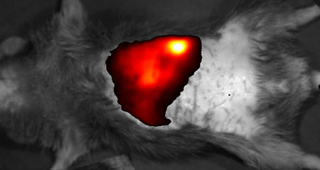
C57BL/6 mouse image taken 1 hour after NIR fluorescent dye injection through the tail vein.
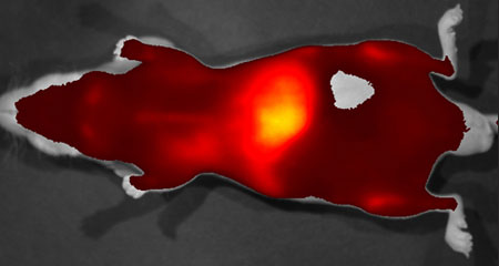
Nude mouse image taken 2 days after Exosome-NIR dye complex injection through the tail vein.
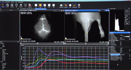
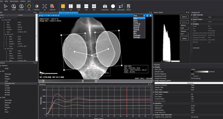
Intelligent in vivo Imaging Viewer & Kinetic Analysis Program
Fast and Convenient Analysis Tools
- One-click analysis
: automatic display of fluorescence level, ROI analysis units, etc. - Image process
: autofluorescence signal removal, merge of multi-spectral images - Report mode
: shows the acquisition setup information, Raw image, ROI, the range of Pseudocolor bar, comments, etc.
Convenient Post-image Analysis and Edition Tools
- *.cif analysis file output
- The display and analysis of four-different images in one screen
- Other supported formats
: tif, bmp, jpg, png
Time-Lapse Imaging and Analysis Software
- Supporting more than 10 analytic algorithms for pharmacokinetics and biodistribution by using the real-time image acquisition
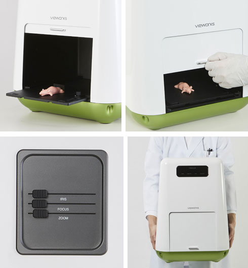
Refined Design & Optional Accessories to Enhance Usability
User-friendly Product Design
Easy Lens Control to Obtain Precise Imaging
- Motorized Iris / Zoom / Focus for detailed adjustments
- Real-time observation of inside of the equipment
- Zoom: 1x - 3x
in vivo Fluorescenct Imaging
- Imaging solid tumors & tracking metastatic tumors
- Assessment of cardiovascular and/or Lymphatic structure and functions
- Evaluating the therapeutic efficacy of new drugs against cancer, arthritis, atherosclerosis, autoimmune disorders or angiogenesis etc.
- Analysis of the pharmacokinetics of new drugs
Pharmacokinetics study of exosomes labeled with NIR fluorescent dyes
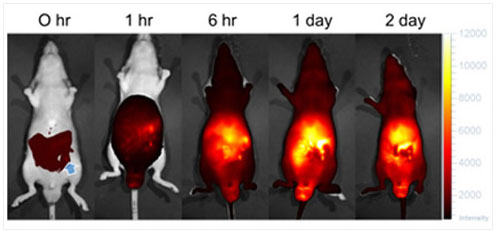
0 hr: immediately after IP injection of the exosome-ICG complex.
The blue arrow indications the injection spot.
Representative Detectable Dyes
| Imaging – Mode | Imaging - Light | Excitation / Emission | Fluorescent Dyes |
|---|---|---|---|
| GFP | Blue | Ex : 390 nm - 490 nm Em : 500 nm - 550 nm |
GFP / EGFP / Alexa 488 / FITC / QD 525 |
| PE | Green | Ex : 530 nm - 570 nm Em : 575 nm - 640 nm |
RFP / DsRed / PE / Alexa 568 / TRITC / QD 585 / QD 605 / QD 625 |
| Cy5.5 | Red | Ex : 620 nm - 650 nm Em : 690 nm - 740 nm |
Cy5.5 / PKE680 / Alexa 680 / Alexa 700 / QD 705 |
| HyperRed | Ex : 630 nm - 680 nm Em : 690 nm - 740 nm |
||
| ICG | NIR | Ex : 740 nm - 790 nm Em : 810 nm - 860 nm |
ICG / QD 800 |
| System | |
|---|---|
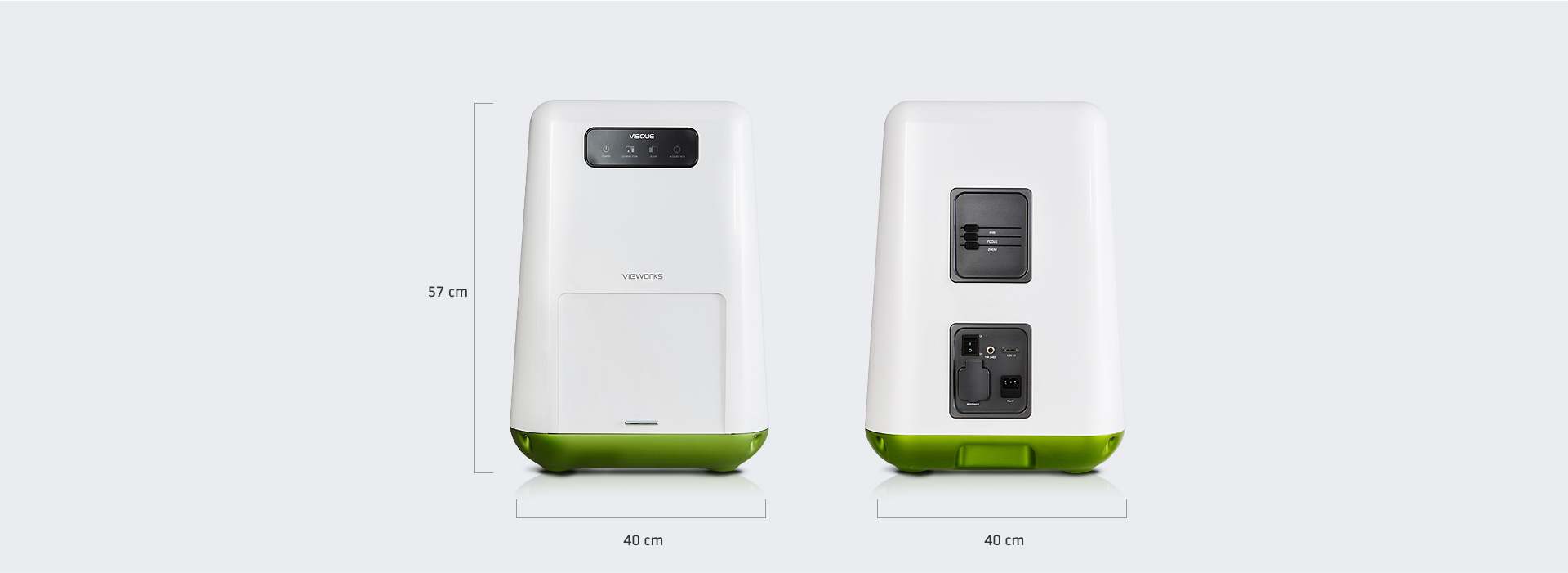
| Dimension | 40 cm x 40 cm x 57 cm |
| Weight | 17 kg |
| Operating Temperature | 10℃ to 27℃ |
| Power | 100 – 240 V AC, 50/60 ㎐, max. 0.5 A at 220 V AC |
| Camera | |
| Sensor | scientific CMOS |
| Resolution (H x V) | 1024 x 1024 |
| Pixel Size | 6.5 um |
| Min. Image Pixel Resolution | 20 um (7.5x) |
| Digital Output | 14 bit |
| Maximum Frame Rate | 30 fps |
| Exposure Time | 0.013s to 3s |
| Detection Spectral Range | 500 ㎚ to 860 ㎚ |
| Interface | USB 3.0 |
| Lens | |
| Control | Zoom / Iris / Focus |
| Zoom (Field of View, H x V) | 15 cm x 15 cm (1x ) ~ 2 cm x 2 cm (7.5x ) |
| Software, CleVueTM | |
| Exclusive File Format | *.CIF (CleVue Image File) Saves all information of an image such as a raw image, analyzed image, ROI information, acquisition information, comments etc. |
| Supported Image File Format | TIFF / Bitmap / JPEG / PNG |
| Image Merging | Merges images of multi-fluorescent dyes |
| Removal of Autofluorescence | Removes autofluorescence or reflection from fluorescent images |
| Report Mode | Displays an analyzed image with color scale bar, analyzed data, acquisition info, comments etc. |
| Kinetics Analysis | • Includes 10 kinds of algorithms, i.e. MTT, BFI, and patented other algorithms to analyze Kinetics • Dynamics graph, i.e. a plot of pixel intensity over time • Map with Kinetics values on an image |
| Excitation Light | |
| Source | LED |
| White Light | epi white LED |
| Emission Filters | |
| Filter Selection | Automated Control |
| Emission Filters | 1 included, 8 optional |
* Specifications are subject to change without prior notice.
* This system is only for research