-

InVivo Smart
소동물 생체 형광 신호 분석 전용 인비보이미징시스템
VISQUE® InVivo Smart
소동물 생체 형광 신호 분석 전용 인비보이미징시스템
고감도 형광 이미지
과학용 카메라
- 하이엔드 과학용 애플리케이션에 최적화된 카메라
- 최소 영상 해상도: 20 µm (7.5x)
고감도 sCMOS 이미지 센서
- 양자 효율(Quantum Efficiency): 72% @ 595 nm
- 다이내믹 레인지: 87 dB
- 암전류(Dark Current): < 10 e-/s/pix @ 30℃
고속 촬영 기능 지원
- 최대 30 fps 속도 촬영 시에도 균일한 영상 품질 유지
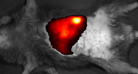
C57BL/6 mouse image taken 1 hour after NIR fluorescent dye injection through the tail vein.
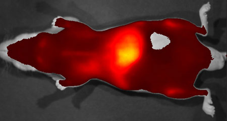
Nude mouse image taken 2 days after Exosome-NIR dye complex injection through the tail vein.
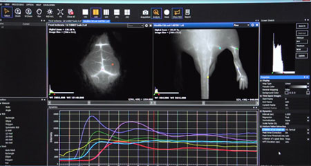
영상 분석 전문 소프트웨어 CleVue™
빠르고 편리한 분석
- 원클릭 분석: 자동 형광레벨 제시, 관심영역(Region of Interest) 분석값 제시 등
쉬운 분석을 지원합니다. - 다중 이미지 분석
: 동시에 최대 4개의 이미지를 비교 분석할 수 있습니다. - 보고서 모드(Report Mode): 한 눈에 다양한 영상 정보(이미지, 관심영역, 촬영조건, 유사 색상 바 범위, 주석 등)를 확인하고 저장할 수 있습니다.
손쉬운 재분석 및 편집
- *.cif 파일: VISQUE 소프트웨어 전용 분석 파일로, 분석에 필요한 정보를
저장하기 때문에 재분석 및 편집을 쉽게 할 수 있습니다. - 기타 지원 포맷
: 이미지 파일(tif, bmp, jpg, png)과 엑셀 파일(csv)을 지원합니다.
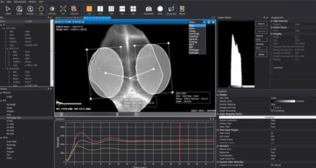
타임랩스(Time-Lapse) 촬영 및 실시간 영상 분석 특화
- 동역학 분석 알고리즘: 실시간 영상을 이용한 생체내 약물동역학(Pharmacokinetics) 및 약물 분포 분석(Biodistribution)에 최적화된 10종류 이상의 알고리즘을 지원합니다.
편의성 향상을 위한 디자인과 옵션
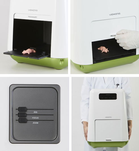
사용자 편의를 고려한 디자인
- 슬라이딩 스테이지 & 스테이지 위치 마커
- 풋 스위치
간편한 렌즈 제어
- 사이드 레버를 통해 실시간 영상을 보며 조절
- 줌 조절 배율: 1x - 7.5x
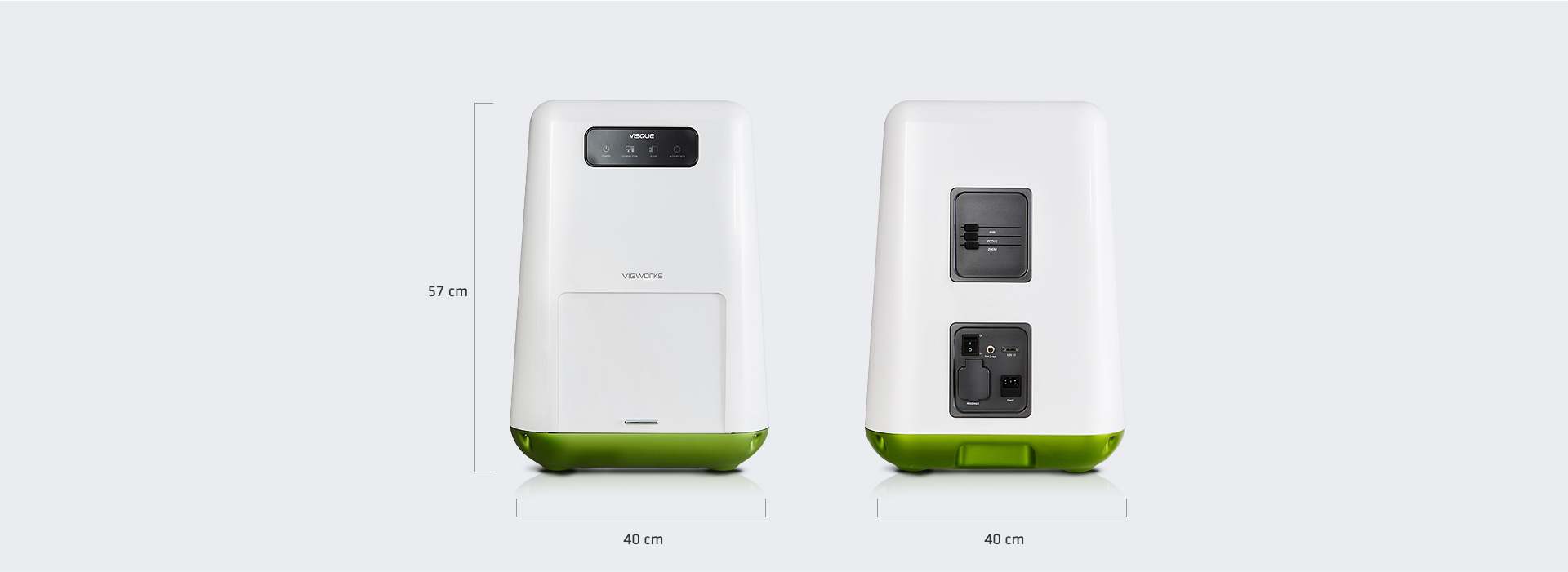
컴팩트한 디자인
- 17kg 내외로 핸드 캐리 가능한 초경량 디자인
- LED 상태 (전원/도어 개방 여부/촬영 모드 등)
형광 신호 영상을 활용한 애플리케이션
- 고형암 영상 및 전이암 위치 추적
- 혈관계, 림프계의 구조 및 기능 평가
- 치료제 효능 평가: 암, 관절염, 동맥경화, 자가면역, 신생혈관 생성 관련 질환
- 타깃 물질의 생체내 약물동역학(Pharmacokinetics) 추적
엑소좀-근적외선 중합체의 생체내 약물동역학 연구
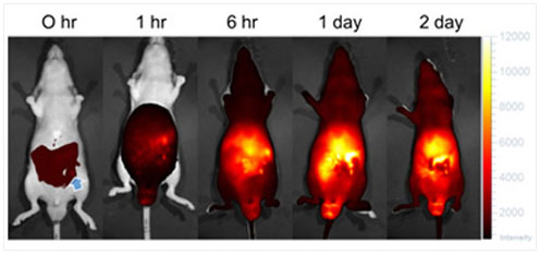
0 hr: 엑소좀-근적외선 중합체 복강내 주사 직후 촬영(파란색 화살표는 주입 위치 표시)
사용 가능 대표 형광
| Imaging – Mode | Imaging - Light | Excitation / Emission | Fluorescent Dyes |
|---|---|---|---|
| GFP | Blue | Ex : 390 nm - 490 nm Em : 500 nm - 550 nm |
GFP / EGFP / Alexa 488 / FITC / QD 525 |
| PE | Green | Ex : 530 nm - 570 nm Em : 575 nm - 640 nm |
RFP / DsRed / PE / Alexa 568 / TRITC / QD 585 / QD 605 / QD 625 |
| Cy5.5 | Red | Ex : 620 nm - 650 nm Em : 690 nm - 740 nm |
Cy5.5 / PKE680 / Alexa 680 / Alexa 700 / QD 705 |
| HyperRed | Ex : 630 nm - 680 nm Em : 690 nm - 740 nm |
||
| ICG | NIR | Ex : 740 nm - 790 nm Em : 810 nm - 860 nm |
ICG / QD 800 |
제품 사양

| System | |
|---|---|
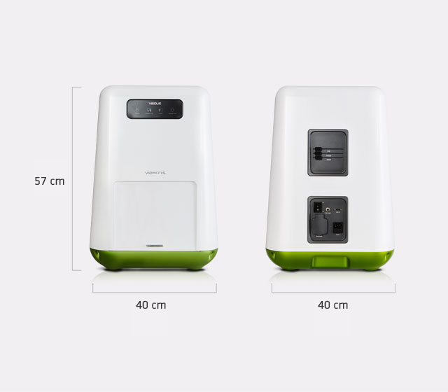
| Dimension | 40 cm x 40 cm x 57 cm |
| Weight | 17 kg |
| Operating Temperature | 10℃ to 27℃ |
| Power | 100 – 240 V AC, 50/60 ㎐, max. 0.5 A at 220 V AC |
| Camera | |
| Sensor | scientific CMOS |
| Resolution (H x V) | 1024 x 1024 |
| Pixel Size | 6.5 um |
| Min. Image Pixel Resolution | 20 um (7.5x) |
| Digital Output | 14 bit |
| Maximum Frame Rate | 30 fps |
| Exposure Time | 0.013s to 3s |
| Detection Spectral Range | 500 ㎚ to 860 ㎚ |
| Interface | USB 3.0 |
| Lens | |
| Control | Zoom / Iris / Focus |
| Zoom (Field of View, H x V) | 15 cm x 15 cm (1x ) ~ 2 cm x 2 cm (7.5x ) |
| Software, CleVueTM | |
| Exclusive File Format | *.CIF (CleVue Image File) Saves all information of an image such as a raw image, analyzed image, ROI information, acquisition information, comments etc. |
| Supported Image File Format | TIFF / Bitmap / JPEG / PNG |
| Image Merging | Merges images of multi-fluorescent dyes |
| Removal of Autofluorescence | Removes autofluorescence or reflection from fluorescent images |
| Report Mode | Displays an analyzed image with color scale bar, analyzed data, acquisition info, comments etc. |
| Kinetics Analysis | • Includes 10 kinds of algorithms, i.e. MTT, BFI, and patented other algorithms to analyze Kinetics • Dynamics graph, i.e. a plot of pixel intensity over time • Map with Kinetics values on an image |
| Excitation Light | |
| Source | LED |
| White Light | epi white LED |
| Emission Filters | |
| Filter Selection | Automated Control |
| Emission Filters | 1 included, 8 optional |
* Specifications are subject to change without prior notice.
* This system is only for research
관련 논문