-

InVivo Smart-LF
紧凑型临床前活体荧光和生物发光成像及分析系统
VISQUE® InVivo Smart-LF
紧凑型临床前活体荧光成像及分析系统
高灵敏度生物发光和荧光成像
采用制冷技术的科研级CMOS相机
- 高端科研应用的更优化解决方案
- 图像最小像素尺寸:26.5μm
- 制冷温度:低于环境温度-50℃
高灵敏度的成像传感器
- 量子效率:
最大. 94%(550 nm 时)
>85%(450 nm – 700 nm 时)
>30%(300 nm – 950 nm 时) - 读出噪声:1.6e-
高速图像采集
- 成像质量均一的图像结果,最快采集速度37fps
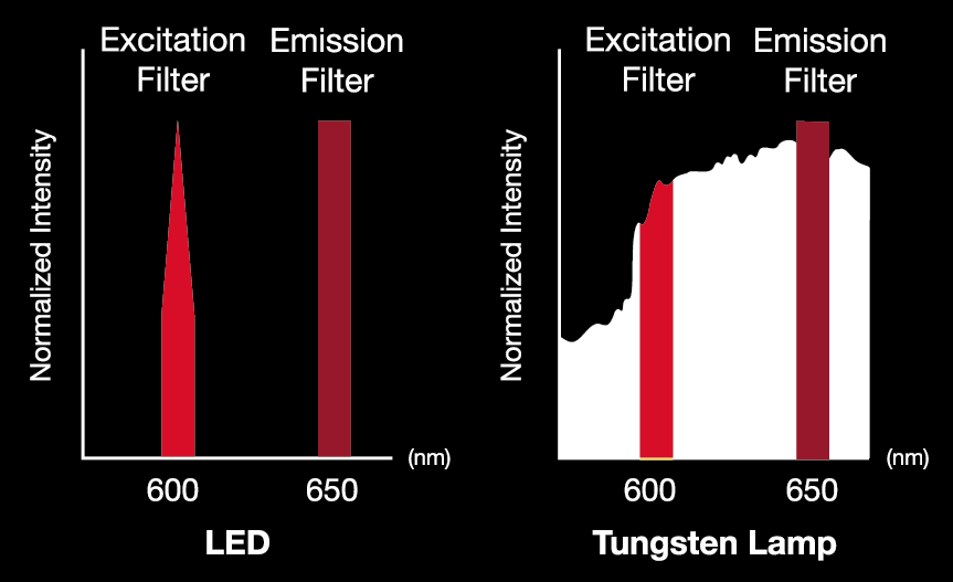
LED光源
- 采用五组LED光源: Blue, Green, Red, HyperRed, N750
- 可针对特定波长范围集中增强光源能量
- 基于LED的光谱分离
- 即刻使用(无需预热/冷却过程)
- 使用寿命长(10,000 - 50,000 小时)
- 背景反射少,信噪比(SNR)高
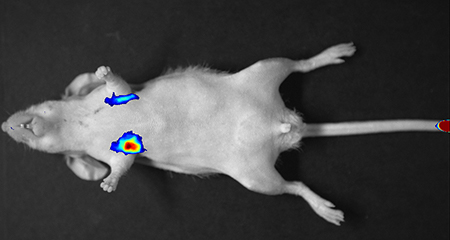
裸鼠尾静脉注射4T1-luc细胞(2.5 x 10⁶)后立刻成像
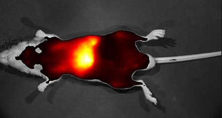
裸鼠尾静脉注射标记了ICG的药物递送材料6小时后成像
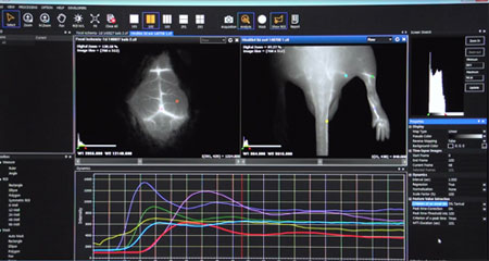
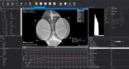
智能活体成像图像查看和动力学分析程序
快速便捷的分析工具
- 键分析
: 自动显示荧光强度,快捷选择ROI区域形状 - 图像处理
: 自发荧光去除,多光谱荧光融合 - 报告模式
: 原始图像,ROI,成像时的设置信息,伪彩条的范围,注解等
便捷的成像后分析和编辑工具
- 输出文件为*.cif
- 最多可同时对比 4 张图像
- 支持多种文件格式
: tif, bmp, jpg, png
专为VISQUE InVivo系列活体成像设计的实时成像和分析软件
- 使用实时成像功能进行药代动力学和生物分布研究,并支持超过10种的动力学分析软法
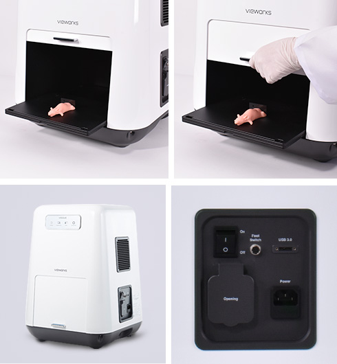
精巧的设计和可选配件增加了设备的适用性
用户友好型产品设计
- 轻巧设计,重量仅22kg,便于挪动
- 紧凑型设计
- 脚踏开关
- 滑动操作台
便捷的镜头设置保证图像质量
- 电动控制镜头,精密调节光圈/焦距/焦点等参数
- 设备内部可实时监控
- 变焦镜头:1-3倍变焦
配件可选,便于操作
- 加热操作台
- 呼吸麻醉机适配器
活体荧光和生物发光成像
- 利用生物发光成像追踪转移性肿瘤
- 利用荧光或生物发光成像进行活体肿瘤成像
- 利用荧光成像评估心血管或淋巴的结构和功能
- 评估新药或新疗法对肿瘤、关节炎、动脉粥样硬化、自身免疫性疾病以及肿瘤新生血管的治疗效果
- 新药或药物递送系统的药代动力学分析
NIR荧光染料标记后的外泌体在体内的药代动力学研究
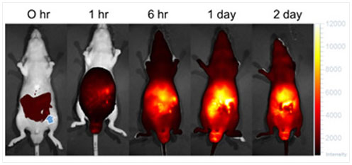
0小时:IP注射外泌体-ICG的复合物后立刻成像
蓝色剪头标注的是注射点
代表性可检测染料
| Imaging – Mode | Imaging - Light | Excitation / Emission | Fluorescent Dyes |
|---|---|---|---|
| GFP | Blue | Ex : 390 nm - 490 nm Em : 500 nm - 550 nm |
GFP / EGFP / Alexa 488 / FITC / QD 525 |
| PE | Green | Ex : 530 nm - 570 nm Em : 575 nm - 640 nm |
RFP / DsRed / PE / Alexa 568 / TRITC / QD 585 / QD 605 / QD 625 |
| Cy5.5 | Red | Ex : 620 nm - 650 nm Em : 690 nm - 740 nm |
Cy5.5 / PKE680 / Alexa 680 / Alexa 700 / QD 705 |
| HyperRed | Ex : 630 nm - 680 nm Em : 690 nm - 740 nm |
||
| ICG | NIR | Ex : 740 nm - 790 nm Em : 810 nm - 860 nm |
ICG / QD 800 |
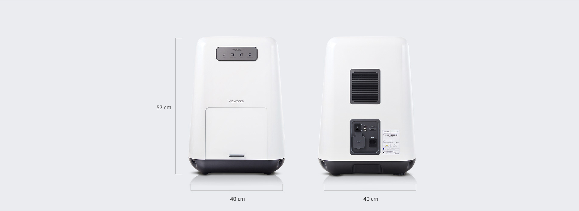
技术规格

| System | |
|---|---|
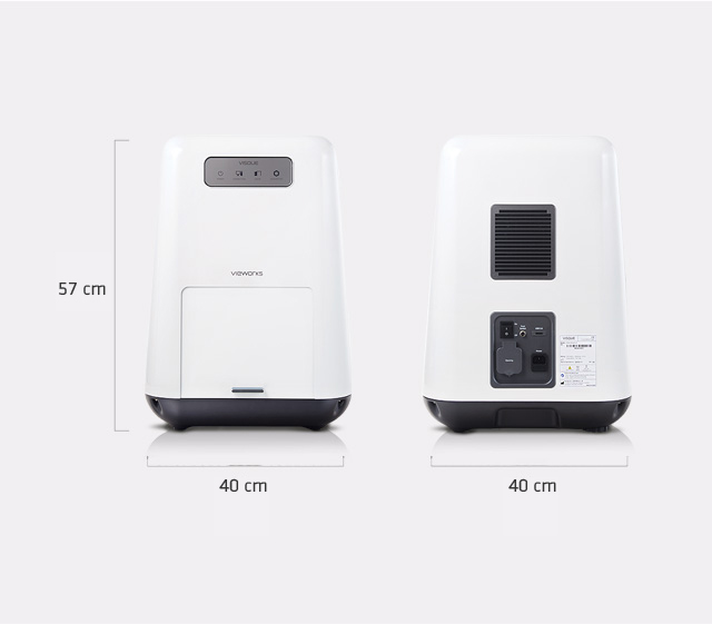
| Dimension | 40 cm x 40 cm x 57 cm |
| Weight | 22 kg (48 lbs) |
| Operating Temperature | 10℃ to 27℃ |
| Power | 100 – 240 V AC, 50/60 ㎐, max. 0.5 A at 220 V AC |
| Camera | |
| Sensor | 1.2” Backside Illuminated sCMOS |
| Resolution (H x V) | 1824 X 1824 |
| Pixel Size | 6.5 um X 6.5 um |
| Min. Image Pixel Resolution | 26.5 um (3x) |
| Digital Output | 16 bit |
| Maximum Frame Rate | 37 fps |
| Exposure Time | 25 ms to 15 min |
| Detection Spectral Range | 220 ㎚ to 940 ㎚ |
| Interface | USB 3.0 |
| Cooling | -20℃, Thermoelectric Peltier Cooling |
| Lens | |
| Control | Motorized Iris / Zoom / Focus |
| Zoom (Field of View, H x V) | 15 cm x 15 cm (1x) to 5 cm X 5 cm (3x) |
| Software, CleVueTM | |
| Supported File Format | cif(exclusive file format) |
| Supported Image File Format | tif, bmp, jpg, png |
| Image Merging | Merges images of multi-fluorescent dyes |
| Removal of Autofluorescence | Removes autofluorescence or reflection from fluorescent images |
| Report Mode | Displays an analyzed image with color scale bar, analyzed data, acquisition info, comments etc. |
| Kinetics Analysis | • Includes 10 kinds of algorithms, i.e. MTT, BFI, and patented other algorithms to analyze Kinetics • Dynamics graph, i.e. a plot of pixel intensity over time • Map with Kinetics values on an image |
| Excitation Light | |
| Source | LED |
| White Light | epi white LED |
| Emission Filters | |
| Filter Selection | Automated Control |
| Emission Filters | Up to 9, optional |
| Stage | |
| Stage Type | Sliding stage, Up to 3 mice |
| Optional Accessory | Heating stage, Anesthesia ventilator adaptor (available in certain regions) |
* Specifications are subject to change without prior notice.
* This system is only for research
引文
下载
文件
目录