-

InVivo ART
Premium Preclinical in vivo Fluorescent & Bioluminescent
Imaging and Analysis System
VISQUE® InVivo ART
VISQUE® InVivo ART is a premium preclinical in vivo fluorescent & bioluminescent imaging and analysis system.
Features
Superior Image Quality & Wide FOV
1) Advanced Cooling Technology
- Camera cooling as low as -100℃
- Adopts TEC technology with additional liquid chiller
2) Back-thinned CCD image sensor
- Quantum Efficiency: 90% @ 450 - 720 nm
- Dark Current: 0.00013@-80℃ (ART 400), 0.00030@ -80℃ (ART 100)
- Pixel well-depth: 100,000 e-
3) High-quality Image
- High resolution image with 13.5 ㎛ (ART 400) / 13 ㎛ (ART 100) pixel size
- Highly sensitive imaging from 350 - 950 ㎚
Full HD Lens with Wider FOV
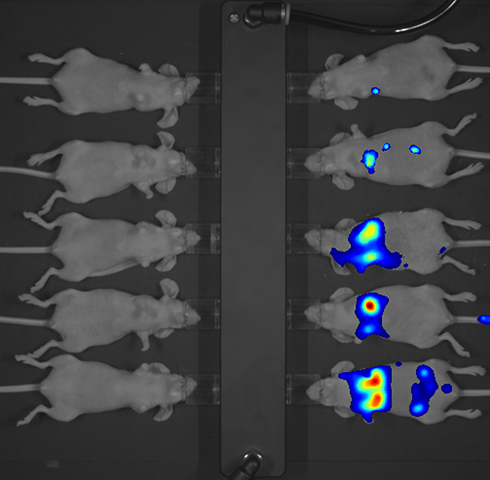
1) 10-mouse FOV
- 27 x 27 cm Field of View (FOV): Sufficient for imaging 10 mice/ 5 rats at a time
Applications
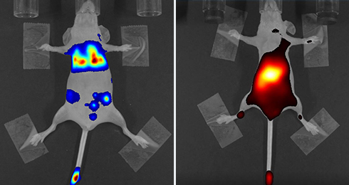
- Small animal in vivo & ex vivo 2D optical imaging
- Tracking tumor metastasis
- Drug biodistribution & transporter targeting
- Pharmacokinetic analysis
- Vascular & lymphatic imaging
- Optical imaging probe development
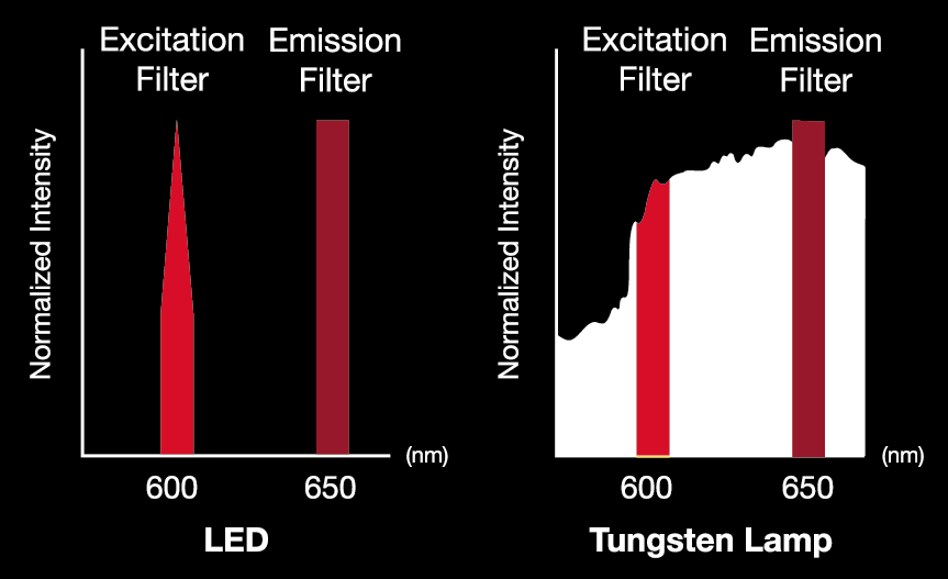
LED light source
- Employs up to 5 LEDs: Blue, Green, Red, HyperRed, NIR
- Strong light power due to concentrated light source energy on a specific wavelength
- LED-based spectral unmixing
- Immediate use (no warm-up/cool-down time)
- Long lifespan (10,000 - 50,000 hours)
- High SNR (signal to noise ratio) due to weak reflection (of excitation light) in the wavelength of emission filter in fluorescence imaing
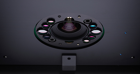
High-performance Fluorescence Filters
1) Emission filters
- Semrock filter: hard-coated filter width up to 6-7 optical density
- Customizable filters (350- 950 nm)
- Undesired reflection removed
2) Excitation filter
- Semrock filter: hard-coated filter width up to 6-7 optical density
- Customizable filters (440 - 770 nm)
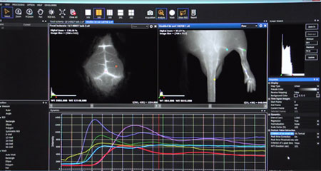
Intelligent in vivo Imaging Viewer & Kinetic Analysis Program
1) Fast and Convenient Analysis Tools
- One-click analysis
: automatic display of fluorescence level and simple setting - Image process
: autofluorescence signal removal, merge of multi-spectral images - Report mode
: the acquisition setup information, raw image, ROI, the range of pseudocolor bar, comments, etc.
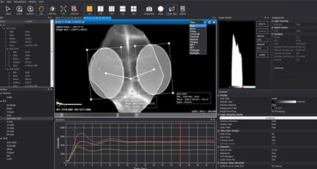
2) Convenient Post-image Analysis and Edition Tools
- *.cif analysis file output
- The display and analysis of four-different images in one screen
- Other supported formats
: tif, bmp, jpg, png
3) Advanced Imaging and Analysis Software
- Time-lapse imaging analysis
- Kinetic analysis: Dynamics graph and 10 kinds of algorithms
- Image analysis:
Autofluorescence removal, Spectral unmixing, Merge of multi-spectral image
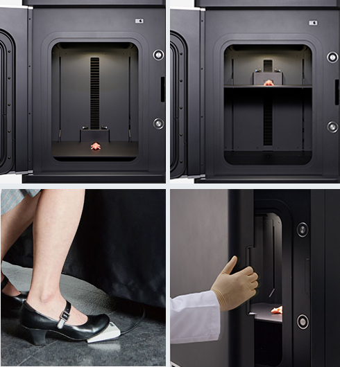
Enhanced Usability with Refined Design & Optional Accessories
1) User-friendly Product Design
- LED status window
- Built-in touch sensor & Electromagnet for easier opening and closing the door
- Interface panel: power switch, foot switch connector, Anesthesia ventilator adaptor outlet
2) Convenient Lens Control to Obtain Precise Imaging
- Convenient and detailed focus control with real-time observation of mice/rats
- Motorized Iris / Zoom / Focus for detailed adjustments
3) Optional Accessories for Convenient Operation
- Black mat
- Foot switch
- Anesthesia ventilator adaptor (availble for limited region)
Representative Detectable Dyes
| Imaging – Mode | Imaging - Light | Excitation / Emission | Fluorescent Dyes |
|---|---|---|---|
| GFP | Blue | Ex : 390 nm - 490 nm Em : 500 nm - 550 nm |
GFP / EGFP / Alexa 488 / FITC / QD 525 |
| PE | Green | Ex : 530 nm - 570 nm Em : 575 nm - 640 nm |
RFP / DsRed / PE / Alexa 568 / TRITC / QD 585 / QD 605 / QD 625 |
| Cy5.5 | Red | Ex : 620 nm - 650 nm Em : 690 nm - 740 nm |
Cy5.5 / PKE680 / Alexa 680 / Alexa 700 / QD 705 |
| HyperRed | Ex : 630 nm - 680 nm Em : 690 nm - 740 nm |
||
| ICG | NIR | Ex : 740 nm - 790 nm Em : 810 nm - 860 nm |
ICG / QD 800 |
* User defined combinations of excitations and emissions are available.
These enables you to detect other fluorescent dyes that are not listed in the table above.
| VISQUE® InVivo ART 400 | VISQUE® InVivo ART 100 | ||
|---|---|---|---|
| System | |||
| Imaging Capability | in vivo imaging, bioluminescence, fluorescence, time-lapse imaging | in vivo imaging, bioluminescence, fluorescence, time-lapse imaging | |
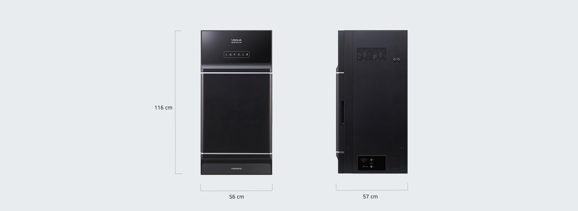
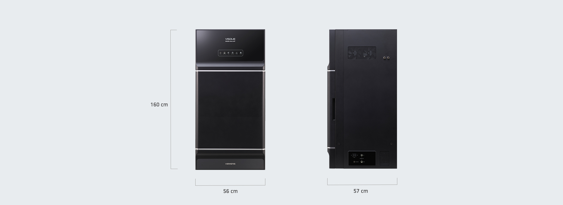
| Weight and Dimension | 56 cm × 57 cm × 116 cm, 140 kg (308.6 lbs) | 56 cm × 57 cm × 116 cm, 140 kg (308.6 lbs) | |
| Camera | |||
| Sensor Size | 1.53” | 0.7” | |
| Sensor | Backside Illuminated CCD, Grade 1 | Backside Illuminated CCD, Grade 1 | |
| Cooling | - 100℃ | - 100℃ | |
| Resolution (H X V) | 2048 x 2048, 4 Mpixel | 1024 × 1024, 1 Mpixel | |
| Pixel Size | 13.5 ㎛ x 13.5 ㎛ | 13 ㎛ x 13 ㎛ | |
| Digital Output | 16 bit | 16 bit | |
| Binning | 1 × 1, 2 × 2, 4 × 4, 8 x 8, 16 x 16 | 1 × 1, 2 × 2, 4 × 4, 8 x 8, 16 x 16 | |
| Fluorescence Filters | |||
| Light Source | LED | LED | |
| Excitation Filter | Customizable filter choices (350 - 950 nm) | Customizable filter choices (350 - 950 nm) | |
| Emission Filters | Customizable filter choices (440 - 770 nm) | Customizable filter choices (440 - 770 nm) | |
| Lens | |||
| Control | Motorized Iris / Zoom / Focus | Motorized Iris / Zoom / Focus | |
| Field of View (FOV) | 27 cm × 27 cm - 10 cm × 10 cm | 27 cm × 27 cm - 8 cm × 8 cm | |
| Stage | |||
| Animal Capacity | 10 mice / 5 rats | 10 mice / 5 rats | |
| Heating Stage | Off / 25℃ / 36℃ | Off / 25℃ / 36℃ | |
| Anesthesia Ventilator Adaptor | Yes | Yes | |
| CleVue Software | |||
| Image Acquisition Mode | Single-frame, Time-lapse imaging | Single-frame, Time-lapse imaging | |
| Supported File Format | cif (exclusive file format), tif, bmp, jpg, png | cif (exclusive file format), tif, bmp, jpg, png | |
| Kinetic Analysis | Dynamics graph and 10 kinds of algorithms for kinetic analysis | Dynamics graph and 10 kinds of algorithms for kinetic analysis | |
| Image Analysis | Quantification, ROI, Autofluorescence removal, Spectral unmixing, Merge of multi-spectral images |
Quantification, ROI, Autofluorescence removal, Spectral unmixing, Merge of multi-spectral images |
|
* Specifications are subject to change without prior notice.
* This system is for research purpose only